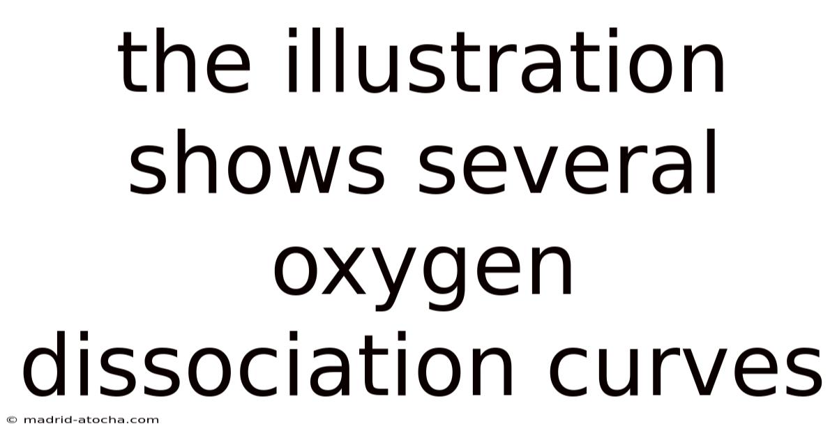
The Illustration Shows Several Oxygen Dissociation Curves
Understanding the Oxygen-Hemoglobin Dissociation Curve: A Window into Blood's Oxygen Transport
The oxygen-hemoglobin dissociation curve is far more than a simple graph; it is a fundamental physiological signature that reveals the intricate dance between oxygen and hemoglobin within our blood. This sigmoid-shaped curve, plotting the percentage of hemoglobin saturated with oxygen against the partial pressure of oxygen (pO₂), is the key to understanding how our bodies efficiently pick up oxygen in the lungs and deliver it to hungry tissues. When an illustration shows several such curves, it is not depicting different species or errors, but rather the dynamic responsiveness of this system. These multiple curves illustrate how physiological conditions—like pH, temperature, carbon dioxide levels, and the presence of other molecules—can shift the curve, dramatically altering hemoglobin's affinity for oxygen. This adaptability is crucial for survival, allowing a single protein to perform optimally in both the oxygen-rich lungs and the oxygen-poor capillaries of active muscles.
The Foundation: The Standard Sigmoid Curve
At its core, the dissociation curve demonstrates hemoglobin's remarkable cooperative binding. The first oxygen molecule binds to hemoglobin with moderate difficulty, but this binding induces a conformational change in the protein's structure, making the binding of subsequent oxygen molecules progressively easier. This is why the curve is S-shaped (sigmoid). At the high pO₂ of the alveoli (around 100 mmHg), hemoglobin becomes almost fully saturated (~97.5%). As blood travels to tissues where pO₂ drops to about 40 mmHg, hemoglobin releases its oxygen cargo, with saturation falling to around 75%. This 22.5% drop represents the oxygen delivered to tissues. The steep middle portion of the curve is physiologically perfect: a small drop in tissue pO₂ results in a large release of oxygen, precisely where it's needed most.
The Shifting Landscape: Factors That Alter the Curve
An illustration showing multiple curves typically highlights how four primary factors cause the curve to shift to the right or left. A right shift means hemoglobin has a lower affinity for oxygen, so it unloads oxygen more readily to tissues. A left shift indicates a higher affinity, meaning hemoglobin holds onto oxygen more tightly, releasing it less easily to tissues.
1. The Bohr Effect: pH and Carbon Dioxide
This is the most significant physiological modulator. Metabolically active tissues produce more carbon dioxide (CO₂) and hydrogen ions (H⁺), lowering pH (making it more acidic).
- Increased CO₂ & Decreased pH (Acidosis): Causes a right shift. CO₂ binds to hemoglobin, forming carbaminohemoglobin, which stabilizes the T (tense) state of hemoglobin with low oxygen affinity. H⁺ ions also promote this T state. The result: oxygen is offloaded more efficiently to active tissues. This is a beautiful feedback loop: working muscles create the conditions that force more oxygen delivery.
- Decreased CO₂ & Increased pH (Alkalosis): Causes a left shift. In the lungs, where CO₂ is being expelled and pH rises, hemoglobin's affinity for oxygen increases, promoting loading.
2. Temperature
- Increased Temperature: Causes a right shift. Active tissues are warmer. Higher temperature reduces hemoglobin's affinity for oxygen, facilitating unloading. Fever can therefore increase oxygen delivery to tissues.
- Decreased Temperature: Causes a left shift. In cooler conditions or peripheral tissues, hemoglobin holds oxygen more tightly.
3. 2,3-Bisphosphoglycerate (2,3-BPG)
This organic phosphate, present in red blood cells, is a critical regulator.
- Increased 2,3-BPG: Causes a right shift. 2,3-BPG binds preferentially to the T state of deoxygenated hemoglobin, stabilizing it and making it harder for oxygen to bind. Levels rise in response to chronic hypoxia (e.g., high altitude, anemia, lung disease), chronic heart failure, and during pregnancy. This adaptation is vital for survival in low-oxygen environments.
- Decreased 2,3-BPG: Causes a left shift. Levels are lower in newborns (especially immediately after birth), in stored blood (a concern for transfusions), and in cases of certain anemias.
4. Carbon Monoxide (CO) Poisoning
This is a special and dangerous case. CO binds to hemoglobin with over 200 times the affinity of oxygen.
- Effect: It causes a left shift of the remaining oxygen-binding sites. Not only does CO occupy heme groups, but its binding also increases the oxygen affinity of the neighboring subunits. This means the hemoglobin that is carrying oxygen is pathological—it clings to it tightly and fails to release it to tissues, causing severe tissue hypoxia. The curve for CO-poisoned blood is not just shifted; it's fundamentally compromised.
Clinical and Physiological Implications of Curve Shifts
The ability to shift the curve is not a laboratory curiosity; it is a matter of life and death.
- Fetal Hemoglobin (HbF): Fetuses have a different form of hemoglobin (HbF) with a left-shifted curve compared to adult hemoglobin (HbA). This higher affinity allows the fetal blood to effectively "steal" oxygen from the maternal circulation across the placenta, where oxygen partial pressures are relatively low.
- Exercise: Working muscles produce heat, CO₂, and lactic acid (lowering pH). All these factors cause a right shift in the local capillary beds, ensuring maximal oxygen unloading precisely where metabolic demand is highest.
- High Altitude Acclimatization: At high altitude, the low pO₂ triggers increased production of 2,3-BPG in red blood cells, shifting the curve right. This compensatory mechanism helps extract the scarce oxygen from the alveoli and deliver it to tissues.
- Anemia and Blood Transfusion: In chronic anemia, 2,3-BPG levels rise (right shift) to
Continuingfrom the point about anemia and transfusion:
-
Anemia and Blood Transfusion: In chronic anemia, 2,3-BPG levels rise (right shift) to enhance oxygen unloading to tissues despite reduced hemoglobin concentration. However, stored blood often has lower 2,3-BPG levels (left shift) compared to fresh blood. Transfusing stored blood can therefore cause a transient left shift in the recipient's hemoglobin, potentially reducing oxygen unloading in tissues and contributing to complications like ischemia, particularly in critically ill patients. This underscores the importance of using fresh or irradiated blood when possible in transfusion medicine.
-
Pregnancy: Fetal hemoglobin (HbF) has a left-shifted oxygen dissociation curve compared to maternal adult hemoglobin (HbA). This higher affinity allows the fetus to effectively "steal" oxygen from the maternal circulation across the placenta, where oxygen partial pressures are relatively low. The maternal blood also undergoes physiological changes, including a mild right shift due to increased 2,3-BPG production, facilitating better oxygen unloading to the placenta and the maternal tissues.
-
Exercise: During physical activity, working muscles generate heat, carbon dioxide (CO₂), and lactic acid, significantly lowering local pH. These factors (increased temperature, decreased pH, increased CO₂) cause a right shift in the oxygen dissociation curve within the capillary beds of active tissues. This shift is crucial, as it dramatically enhances the unloading of oxygen from hemoglobin to the rapidly metabolizing cells, meeting the increased oxygen demand.
-
High Altitude Acclimatization: Exposure to high altitude results in chronic hypoxia (low arterial pO₂). The body responds by increasing red blood cell production (polycythemia) and, critically, by elevating 2,3-BPG levels within red blood cells. This rise in 2,3-BPG causes a right shift in the oxygen dissociation curve. The right shift improves oxygen unloading from hemoglobin in the tissues, compensating for the lower arterial oxygen saturation and helping to maintain adequate tissue oxygen delivery despite the reduced partial pressure gradient.
Conclusion:
The oxygen dissociation curve is a dynamic and exquisitely regulated entity, reflecting the body's remarkable ability to adapt hemoglobin's oxygen affinity to meet changing physiological demands. Factors like temperature, pH, CO₂, 2,3-BPG, fetal hemoglobin, and even the presence of carbon monoxide profoundly influence the curve's position. A right shift enhances oxygen unloading to tissues, vital during exercise, at high altitude, or in states of increased metabolic demand or acidosis. Conversely, a left shift increases oxygen affinity, facilitating oxygen loading in the lungs and protecting tissues in conditions like fetal development or when oxygen availability is critically low. Understanding these shifts is fundamental to physiology and medicine, explaining adaptations to hypoxia, the challenges of transfusion, the risks of carbon monoxide poisoning, and the intricate balance the body maintains to ensure adequate oxygen delivery to every cell. This constant modulation of hemoglobin's oxygen-binding properties is a cornerstone of life.
Latest Posts
Latest Posts
-
Anastasio Pereira No Esta Convencido De La Invitacion De Barreto
Mar 28, 2026
-
On The Number Line Below Length Ab
Mar 28, 2026
-
Your Health Today Choices In A Changing Society
Mar 28, 2026
-
4 7 8 How Many Players In The Game
Mar 28, 2026
-
Excel Project Historical Financial Statements Assignment
Mar 28, 2026