A Ribbon Diagram Of A Zinc Metallo-beta-lactamase Protein Is Shown
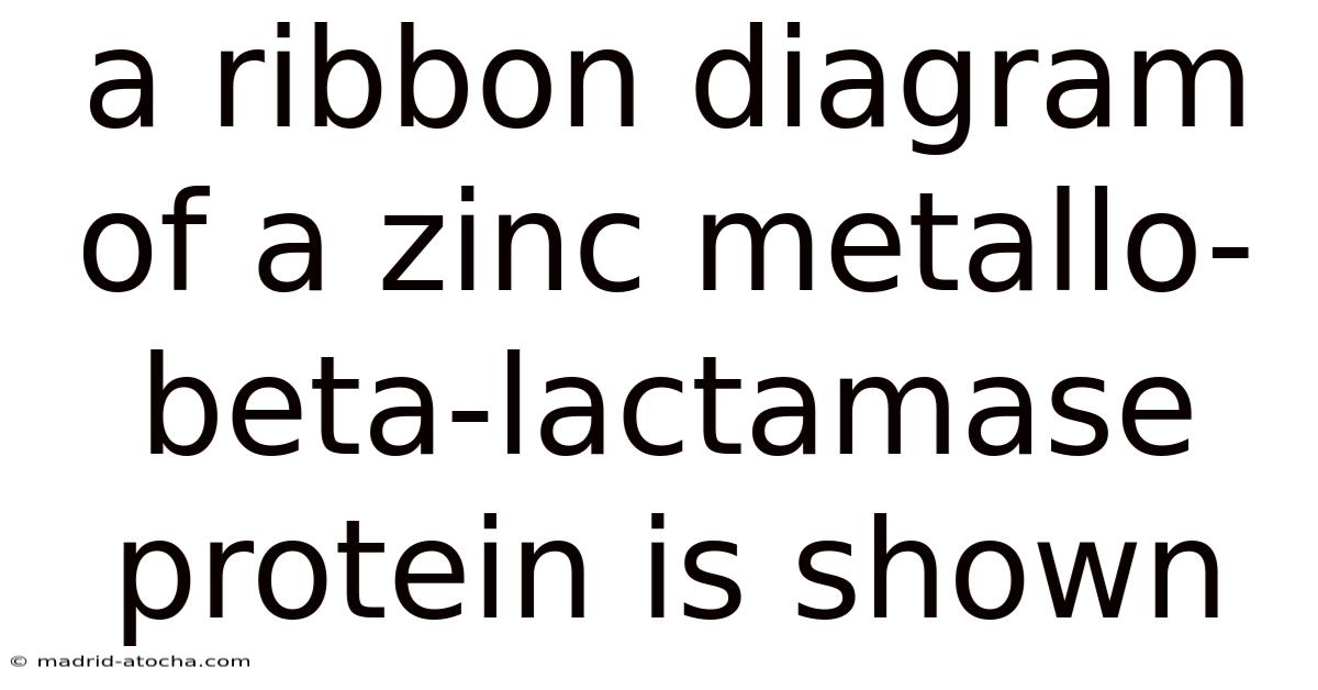
A ribbon diagram of a zinc metallo-beta-lactamase protein is shown. This three-dimensional representation highlights the structural features of the enzyme, which plays a critical role in the breakdown of beta-lactam antibiotics. The ribbon diagram, a widely used visualization tool in structural biology, simplifies the complex arrangement of amino acids into a continuous line, making it easier to analyze the protein’s conformation and functional domains. The zinc metallo-beta-lactamase, a member of the metalloenzyme family, is particularly significant due to its ability to hydrolyze beta-lactam antibiotics, rendering them ineffective. Understanding the structure of this enzyme through such diagrams is essential for developing strategies to combat antibiotic resistance.
The zinc metallo-beta-lactamase protein is a key player in the mechanism of antibiotic resistance. These enzymes, which require zinc ions for their activity, cleave the beta-lactam ring of antibiotics like penicillins and cephalosporins, preventing them from inhibiting bacterial cell wall synthesis. The ribbon diagram provides a visual framework to study how the enzyme’s structure facilitates this process. By examining the spatial arrangement of amino acids and the positioning of zinc ions, researchers can gain insights into the catalytic mechanism and identify potential targets for drug development.
The structure of the zinc metallo-beta-lactamase is characterized by a beta-barrel fold, a common motif in many enzymes. This fold consists of multiple beta-strands arranged in a cylindrical structure, creating a hydrophobic interior that stabilizes the protein. The ribbon diagram illustrates this fold, showing how the beta-strands are connected and how they contribute to the overall stability of the enzyme. The zinc ions, which are essential for the enzyme’s activity, are typically located in the active site, where they coordinate with specific amino acid residues to facilitate the hydrolysis of the beta-lactam ring.
In the ribbon diagram, the active site is often depicted as a cleft or pocket within the protein structure. This region is critical for the enzyme’s function, as it contains the zinc ions and the amino acids that interact with the substrate. The diagram allows researchers to visualize how the active site is structured and how it interacts with the antibiotic molecules. For example, the zinc ions may be coordinated by histidine, cysteine, and glutamate residues, which form a tight binding pocket that stabilizes the zinc and positions it for catalytic activity.
The catalytic mechanism of the zinc metallo-beta-lactamase is a complex process that involves the coordination of zinc ions with the substrate. The ribbon diagram helps to illustrate how the enzyme’s structure enables this mechanism. The zinc ions act as Lewis acids, polarizing the carbonyl group of the beta-lactam ring and making it more susceptible to nucleophilic attack. This process is facilitated by the precise arrangement of amino acids in the active site, which ensures that the zinc ions are positioned correctly for catalysis. The diagram also highlights the role of water molecules, which may act as a nucleophile in the hydrolysis reaction.
The significance of the zinc metallo-beta-lactamase structure extends beyond its role in antibiotic resistance. The ribbon diagram provides a foundation for understanding how the enzyme’s structure influences its function and how it can be targeted by inhibitors. By analyzing the spatial relationships between amino acids and zinc ions, researchers can design molecules that bind to the active site and block the enzyme’s activity. This approach is crucial for developing new antibiotics that can overcome resistance mechanisms.
The ribbon diagram also reveals the evolutionary relationships between different metallo-beta-lactamases. By comparing the structures of various enzymes, scientists can identify conserved regions that are essential for function and regions that vary between species. This information is valuable for understanding the diversity of these enzymes and for predicting how they might evolve in response to environmental pressures. The diagram serves as a tool for studying the structural basis of antibiotic resistance and for identifying potential targets for therapeutic intervention.
In addition to its role in antibiotic resistance, the zinc metallo-beta-lactamase structure has implications for other areas of biochemistry. The enzyme’s ability to hydrolyze beta-lactam rings is a unique feature that has inspired the development of synthetic analogs and inhibitors. The ribbon diagram allows researchers to explore these possibilities by providing a detailed view of the enzyme’s active site and the interactions that occur during catalysis. This knowledge is essential for advancing the field of enzyme engineering and for creating new therapeutic agents.
The study of zinc metallo-beta-lactamase through ribbon diagrams also contributes to the broader understanding of protein structure and function. By visualizing the three-dimensional arrangement of amino acids, scientists can gain insights into how proteins fold and how their structures determine their functions. This knowledge is fundamental to the field of structural biology and has applications in various areas, including drug discovery, enzyme engineering, and the study of protein dynamics.
The ribbon diagram of the zinc metallo-beta-lactamase protein is a powerful tool for researchers studying antibiotic resistance and enzyme function. It provides a clear and detailed representation of the enzyme’s structure, highlighting the critical regions involved in catalysis and resistance. By analyzing this diagram, scientists can better understand the mechanisms by which the enzyme operates and how it can be targeted for therapeutic purposes. The insights gained from such studies are essential for addressing the growing challenge of antibiotic resistance and for developing new strategies to combat bacterial infections.
In conclusion, the ribbon diagram of the zinc metallo-beta-lactamase protein offers a valuable perspective on the structural and functional aspects of this important enzyme. By visualizing the enzyme’s three-dimensional structure, researchers can gain insights into its catalytic mechanism, its role in antibiotic resistance, and its potential as a target for drug development. The diagram not only enhances our understanding of the enzyme’s function but also provides a foundation for future research aimed at overcoming resistance and improving treatment options. As the field of structural biology continues to advance, the ribbon diagram remains an essential tool for exploring the complexities
of biological systems and for driving innovation in medicine and biotechnology. The ability to visualize and manipulate these complex structures at the molecular level promises to unlock new avenues for tackling global health challenges, and the zinc metallo-beta-lactamase ribbon diagram serves as a compelling example of this potential. Further refinement of these diagrams, incorporating dynamic data and computational modeling, will undoubtedly lead to even deeper insights and more effective therapeutic strategies in the years to come. Ultimately, the continued exploration of protein structures like this one is crucial for safeguarding public health and ensuring the efficacy of antimicrobial treatments in a world increasingly threatened by resistant pathogens.
Building on this understanding, researchers are now focusing on integrating advanced imaging techniques like cryo-electron microscopy and X-ray crystallography to further refine these structural models. These methods allow for higher-resolution views of proteins in various states, including active and inactive conformations, offering a more nuanced picture of how proteins interact with substrates and inhibitors. Such detailed insights are crucial for designing precise inhibitors that can disrupt the enzyme’s function without causing unintended side effects.
Moreover, the study of protein structures is increasingly interdisciplinary, merging computational biology with experimental validation. Machine learning algorithms are now being employed to predict protein folding patterns and identify potential binding sites, accelerating the discovery of new therapeutics. This synergy between technology and biology is reshaping the landscape of biomedical research, enabling scientists to tackle complex challenges with greater precision.
As we continue to unravel the intricacies of protein architecture, the implications extend beyond individual molecules. They inform broader strategies in personalized medicine, where understanding a patient’s unique protein profile can guide tailored treatments. This evolving narrative underscores the transformative power of structural biology in addressing some of the most pressing issues in healthcare.
In summary, the journey from visualizing protein structures to applying this knowledge in real-world solutions highlights the dynamic nature of scientific discovery. Each advancement not only deepens our biological understanding but also empowers innovation in addressing health crises. The future of this field lies in its ability to bridge the gap between molecular insights and practical applications, ensuring that scientific progress keeps pace with the needs of society.
Concluding, the exploration of protein structures remains a cornerstone of modern science, driving breakthroughs that enhance our capacity to combat disease and improve quality of life. The ongoing efforts to decode these intricate frameworks are not just academic exercises—they represent a vital step toward a healthier tomorrow.
Latest Posts
Latest Posts
-
Simon Companys Year End Balance Sheets Follow
Mar 27, 2026
-
Troy Engines Limited Manufactures A Variety
Mar 27, 2026
-
Which Solution Below Has The Highest Concentration Of Hydronium Ions
Mar 27, 2026
-
Blue And Yellow Streams Of Paint At 60
Mar 27, 2026
-
Which Of Mcdonalds Peers Offers The Best Relative
Mar 27, 2026